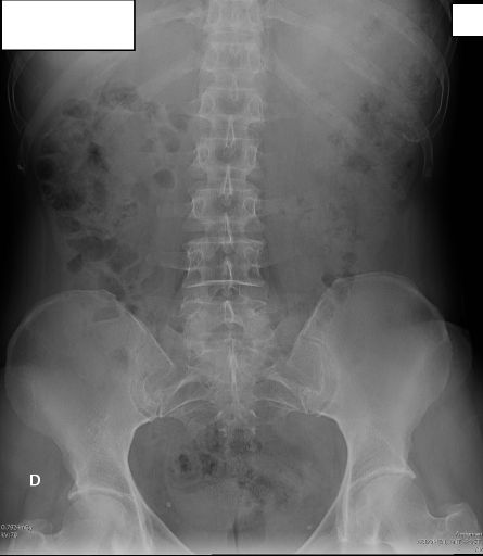
AP ABDOMEN
Anteroposterior supine projection • KUB radiography (Kidneys, Ureters, Bladder)

MAIN INDICATION AND CLINICAL UTILITY
Pathologies demonstrated: Abnormal masses, abnormal intestinal gas patterns, and calculi (stones).
OBTAIN BEFORE CONTRAST STUDIES OR WHEN CLINICALLY INDICATED
Also known as "KUB" (Kidneys, Ureters, Bladder) or "plain abdominal film".
Exposure Factors
Medium exposure: Parameters for optimal visualization of abdominal structures
Anatomical Structures Visible
Should be clearly observed:
- Lower thoracic vertebrae (T12-L1) and upper sacrum
- Abdominal cavity from diaphragm to pelvis
- Psoas muscles bilaterally (if patient not too obese)
- Intestinal gas pattern throughout abdomen
- Renal shadows and bladder outline if distended
Cassette Size and Orientation
Longitudinal orientation to cover from diaphragm to pelvis
Patient Positioning
Central Ray Point
Direction: Perpendicular to center of cassette
Location: Approximately at level of iliac crests for adequate coverage
Special Patient Considerations
Obese Patients
Increase kV and mAs according to thickness adjustment chart. Ensure complete inclusion of abdomen.
Acutely Ill Patients
If patient cannot lie supine, obtain upright abdomen projection first for air-fluid levels.
Pediatric Patients
Reduce exposure according to age and ALARA principles. Use gonadal shielding when appropriate.
CRITICAL NOTE FOR WOMEN OF CHILDBEARING AGE
Always document last menstrual period date before exposure.
Follow the "10-day rule": Perform only during first 10 days after onset of menstruation when pregnancy is unlikely.
Apply gonadal shielding when possible without obscuring diagnostic area
Patient Instructions
"Take a deep breath in, hold it, and remain completely still during the exposure"
Maintain position without movement and apnea during radiographic exposure
Specific Findings to Look For
Bowel Gas Pattern
Distribution and caliber of intestinal gas
Calculi
Renal, ureteral or bladder stones
Masses
Abnormal soft tissue densities
Psoas Shadows
Bilateral visualization of psoas muscles
Common Technical Challenges
Frequent problems in AP abdomen projection:
- Inadequate coverage excluding diaphragm or pelvis
- Patient rotation causing asymmetry of abdominal structures
- Inadequate exposure for obese patients
- Respiratory motion blurring diaphragmatic outlines
Solution: Ensure full inspiration breath-hold and center midway between xiphoid and symphysis